Share this
Coordination Structure of Samarium Diiodide in a Tetrahydrofuran–Water Mixture
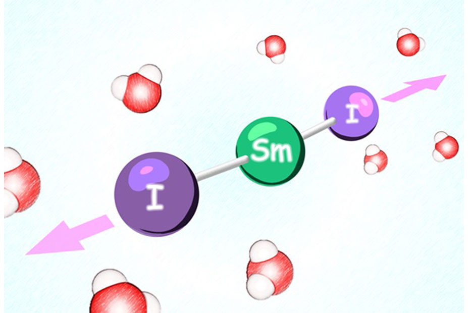
Coordination Structure of Samarium Diiodide in a Tetrahydrofuran–Water Mixture
2023/02/03
Successful development of next-generation crystalline sponge - Expected to be applied to fields such as drug discovery as a new structural analysis method -
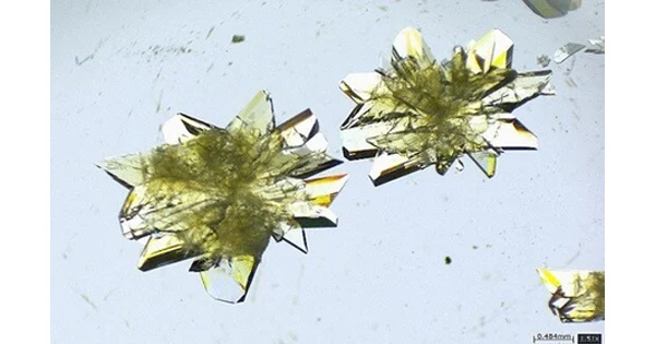
Successful development of next-generation crystalline sponge - Expected to be applied to fields such as drug discovery as a new structural analysis method -
2024/11/06
Proximity effect of emergent field from spin ice in an oxide heterostructure
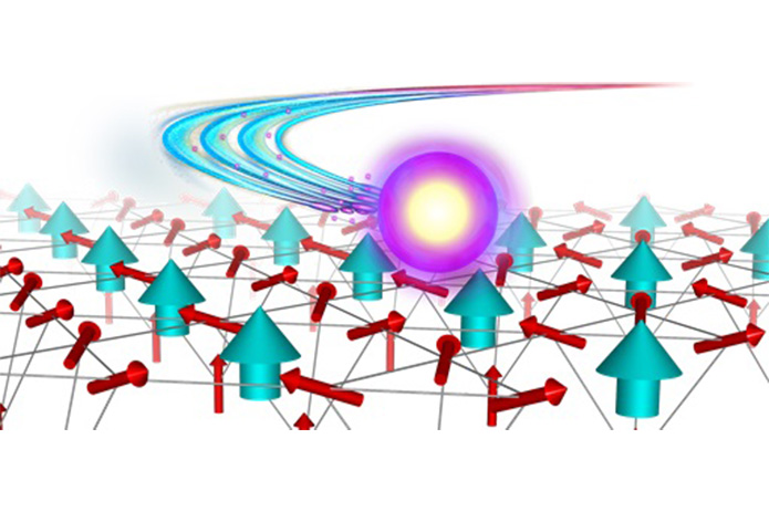
Proximity effect of emergent field from spin ice in an oxide heterostructure
2024/03/14