Share this
Multicolor Activatable Raman Probes for Simultaneous Detection of Plural Enzyme Activities
![]()
Multicolor Activatable Raman Probes for Simultaneous Detection of Plural Enzyme Activities
2020/11/25
Optical Activity in Achiral Crystals: Challenging a Fundamental Principle
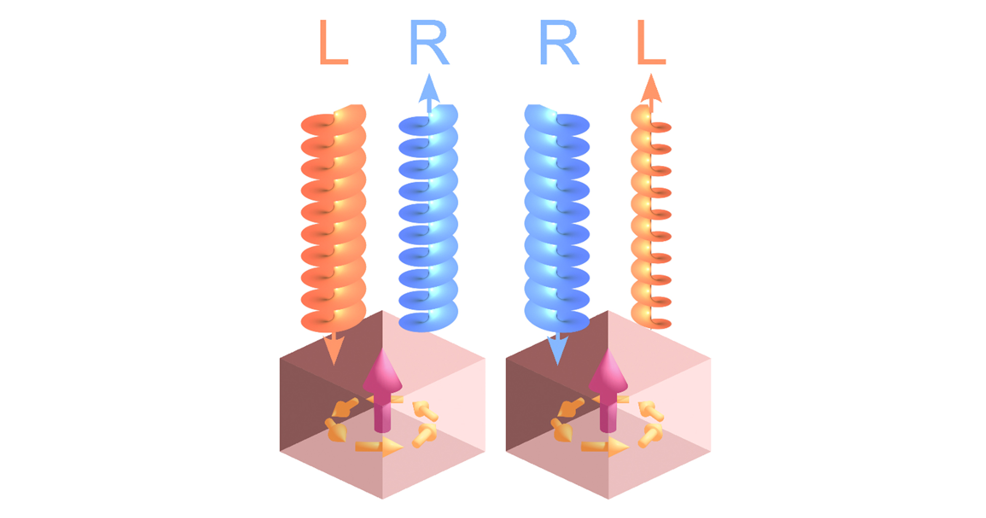
Optical Activity in Achiral Crystals: Challenging a Fundamental Principle
2026/05/21
Isolating single Euglena gracilis cells by glass microfluidic for Raman analysis of paramylon biogenesis
![]()
Isolating single Euglena gracilis cells by glass microfluidic for Raman analysis of paramylon biogenesis
2019/07/09