PRESS RELEASE
- Research
- 2018
A microfluidic device for isolating intact chromosomes from single mammalian cells and probing their folding stability by controlling solution conditions
Authors
Tomohiro Takahashi, Kennedy O. Okeyo, Jun Ueda, Kazuo Yamagata, Masao Washizu & Hidehiro Oana
Abstract
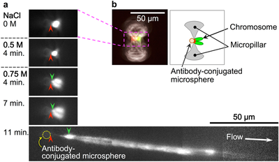
Chromatin folding shows spatio-temporal fluctuations in living undifferentiated cells, but fixed spatial heterogeneity in differentiated cells. However, little is known about variation in folding stability along the chromatin fibres during differentiation. In addition, effective methods to investigate folding stability at the single cell level are lacking. In the present study, we developed a microfluidic device that enables non-destructive isolation of chromosomes from single mammalian cells as well as real-time microscopic monitoring of the partial unfolding and stretching of individual chromosomes with increasing salt concentrations under a gentle flow. Using this device, we compared the folding stability of chromosomes between non-differentiated and differentiated cells and found that the salt concentration which induces the chromosome unfolding was lower (≤500 mM NaCl) for chromosomes derived from undifferentiated cells, suggesting that the chromatin folding stability of these cells is lower than that of differentiated cells. In addition, individual unfolded chromosomes, i.e., chromatin fibres, were stretched to 150–800 µm non-destructively under 750 mM NaCl and showed distributions of highly/less folded regions along the fibres. Thus, our technique can provide insights into the aspects of chromatin folding that influence the epigenetic control of cell differentiation.
Scientific Reports: www.nature.com/articles/s41598-018-31975-5