PRESS RELEASE
- Research
- 2022
Millimetre-scale magnetocardiography of living rats with thoracotomy
Authors
Keigo Arai, Akihiro Kuwahata, Daisuke Nishitani, Ikuya Fujisaki, Ryoma Matsuki, Yuki Nishio, Zonghao Xin, Xinyu Cao, Yuji Hatano, Shinobu Onoda, Chikara Shinei, Masashi Miyakawa, Takashi Taniguchi, Masatoshi Yamazaki, Tokuyuki Teraji, Takeshi Ohshima, Mutsuko Hatano, Masaki Sekino, and Takayuki Iwasaki
Abstract
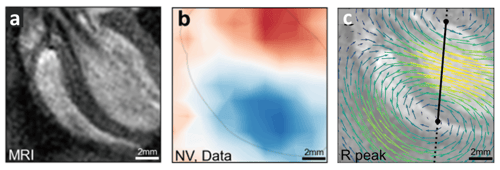
Magnetocardiography is a contactless imaging modality for electric current propagation in the cardiovascular system. Although conventional sensors provide sufficiently high sensitivity, their spatial resolution is limited to a centimetre-scale, which is inadequate for revealing the intra-cardiac electrodynamics such as rotational waves associated with ventricular arrhythmias. Here, we demonstrate invasive magnetocardiography of living rats at a millimetre-scale using a quantum sensor based on nitrogen-vacancy centres in diamond. The acquired magnetic images indicate that the cardiac signal source is well explained by vertically distributed current dipoles, pointing from the right atrium base via the Purkinje fibre bundle to the left ventricular apex. We also find that this observation is consistent with and complementary to an alternative picture of electric current density distribution calculated with a stream function method. Our technique will enable the study of the origin and progression of various cardiac arrhythmias, including flutter, fibrillation, and tachycardia.
(K. Arai et al. Communications Physics)
Communications Physics: https://www.nature.com/articles/s42005-022-00978-0